How Microscopes Reveal Tiny Body Structures Like Airway Cilia
5 min read / 2026-09-18
Microscopes magnify structures far too small to see with the naked eye, which is how doctors and scientists can watch cilia beating inside a child's airway to diagnose rare diseases.
What it means
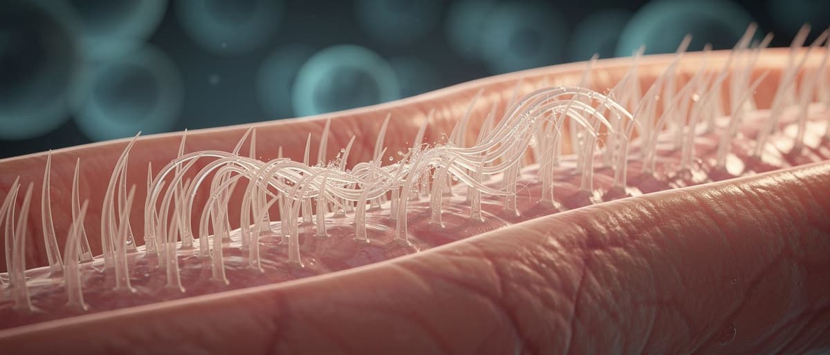
A microscope is a tool that magnifies, or enlarges, things that are too small for our eyes to see on their own. The human eye can normally spot objects down to about 0.1 millimeters, roughly the width of a thin hair. Cilia, the hair-like structures that beat inside airways, are far smaller than that, so scientists rely on microscopes to study them at all, let alone watch them move in real time.
How it works
Light microscopes use glass lenses to bend and focus light, making small objects appear larger, usually up to about 1,000 times their real size. Electron microscopes use beams of electrons instead of light and can magnify objects millions of times, revealing even finer detail like the internal structure of a single cell. For watching movement, such as cilia beating 10 to 15 times per second, scientists often pair a light microscope with a high-speed camera, since electron microscopes usually require samples to be still.
A simple example
Imagine trying to read a text message from across a football field. You cannot see the letters at all until you use binoculars to bring them closer. A microscope does the same job for biology: a doctor cannot see cilia beating with the naked eye, but placing a tissue sample under a microscope and recording it on video lets them watch the tiny hairs move in real time, almost like slowing down a fast cricket bowl to study the wrist action frame by frame.
Why people talk about it
Microscopy videos have become a way to make invisible biology understandable to everyone, not just scientists, which is why contests exist to celebrate striking footage. In medicine, video microscopy matters because some conditions, like the rare genetic disease behind faulty cilia, do not show up on routine chest X-rays or standard breathing tests. Only by directly watching the cilia's motion can doctors confirm whether the tiny hairs are beating in the coordinated wave pattern needed to clear mucus and germs.
What to remember
Magnification alone is not enough; resolution, or the ability to distinguish fine detail, and in some cases speed of recording, are equally important depending on what a scientist wants to observe. Light microscopes are cheaper, faster, and better for watching motion, while electron microscopes offer far higher magnification but usually only for still samples. Choosing the right microscope depends entirely on the question being asked, whether it is counting bacteria, studying a virus, or diagnosing a breathing disorder in a child.
Key words
Magnification
How much larger an object appears through a microscope compared to its actual size.
Resolution
A microscope's ability to show fine detail clearly, separate from how large the image appears.
Electron microscope
A microscope that uses beams of electrons instead of light to achieve extremely high magnification.
Cilia
Tiny hair-like structures on cells that beat in coordinated waves to move mucus and particles.
Key facts
- 1Light microscopes typically magnify objects up to about 1,000 times their real size using bent light and glass lenses.
- 2Electron microscopes use electron beams instead of light and can magnify objects millions of times for finer detail.
- 3The human eye alone usually cannot resolve objects smaller than about 0.1 millimeters, roughly the width of a human hair.
- 4High-speed cameras attached to light microscopes let scientists record fast movements, such as cilia beating 10 to 15 times per second.
- 5Video microscopy can catch movement disorders that standard medical tests like X-rays or breathing tests often miss.
Why it matters
Understanding how microscopes work explains why doctors needed video footage, not just a photo or X-ray, to diagnose the child's rare ciliary disease in the news story.
Sources
- National Institutes of Health
- BBC News