How heart valve collagen survives decades of beating and enzymes
5 min read / 2026-10-15
Heart valve flaps contain collagen, yet the fluid around the heart holds enzymes that break collagen down. Pig heart tests suggest the pattern of stretch in each heartbeat helps protect it.
The puzzle: strong collagen next to collagen-cutting enzymes
Collagen is a protein that keeps heart valve flaps, called leaflets, strong. The flaps open to let blood move in one direction and then shut to stop it flowing back, millions of times every year. The fluid around the heart also contains collagenases, enzymes designed to break collagen down. Both facts are true at once, so the question is why healthy valves still stay sturdy for decades.
Strain ratio: stretch is not just how much, but which way
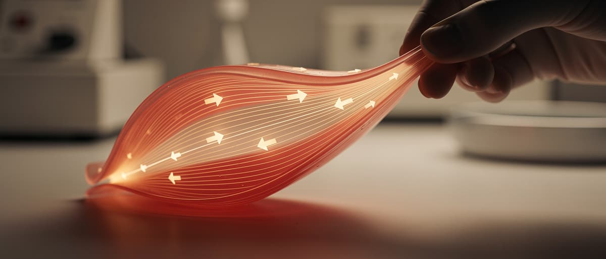
Strain describes how much a tissue is stretched. A valve flap is pulled in more than one direction at each beat, so researchers can describe the stretch as a ratio. In a healthy heart with the aortic valve open, the flap is stretched to 70% of its maximum lengthwise and only 20% of its maximum sideways. The study treated this lengthwise-to-sideways pattern as the key feature, not only the overall amount of stretching.
Suppose the same pattern is shrunk or changed
Suppose a flap is stretched 70% lengthwise and 20% sideways. Another piece is stretched 35% and 10%. The amounts are smaller, but the ratio is the same. The researchers ran tests like this, and collagen stayed undamaged even with higher enzyme levels. Now suppose a piece is stretched equally in both directions. In the tests, changing the ratio like this led to collagen breakdown, whatever the enzyme level was.
What the tests did and what they link to disease
The team used pig hearts. They varied the strain on the flaps, the direction of the pull, and the collagenase level in the surrounding fluid. Afterwards they examined the microstructure of the tissue to see whether it was intact or damaged. The authors say stretching in the same direction as the collagen-rich fibers acts as a protective mechanism while the natural ratio is kept. They suggest that heart disease, by altering strain, may remove this defense, which could explain why flaps degrade quickly in disease.
Limits: pig tissue, one mechanism, a proposed use
The measured result comes from pig hearts, so it is not a test in people. Earlier work found a similar protective effect in tendons, but only pulled tissue in one direction. Valve flaps are stretched in more than one direction, which makes this finding a step forward. The authors also say the work may help engineers design new biomaterials for replacement valves. That is a suggested use, not a product in use.
Key words
Collagen
A strong protein that gives tissues such as heart valve flaps their strength.
Collagenase
An enzyme that breaks collagen down.
Leaflet
A flap of tissue in a heart valve that opens to let blood pass one way and shuts to stop backflow.
Strain
A measure of how much a material is stretched compared with its maximum.
Key facts
- 1Collagen keeps heart valve flaps strong, but the fluid around the heart contains collagenases, enzymes designed to break collagen down.
- 2In a healthy open aortic valve, the flap is stretched to 70% of its maximum lengthwise and 20% sideways.
- 3The tests also used other strain levels with the same ratio, such as 35% and 10%, and collagen stayed undamaged even with higher enzyme levels.
- 4When the strain ratio was changed, including equal stretch in both directions, collagen broke down regardless of collagenase level.
- 5The experiments used pig hearts, and the suggested application is engineering new biomaterials for replacement heart valves.
Why it matters
It offers a mechanical explanation for why healthy valves last and why they may decay faster when disease changes how they stretch. Replacement valve design is only a proposed use.
Sources
- Adapted from Futurity: How do heart valves last so long? · NC State · Source license